
Is a lump in the liver always a liver cancer? Gd-EOB-DTPA enhanced MRI will speak the truth
Is a lump in the liver always a liver cancer? It depends. Tumor can be benign and malignant. Most lumps found in the liver are malignant lesions, primary liver cancer and cholangiocarcinoma for example. And there are also benign lesions, such as Hepatic hemangioma, hepatic adenoma, hepatic focal nodular hyperplasia(FNH). When a lump is detected, the first step is to identify its nature.
In recent years, Hepatocellular Carcinoma (HCC) sufferers are mounting up in different parts of the world and a high percentage of victims died from it, posing a threat to health care services. The early diagnosis of HCC, therefore, has been high on the agenda. In this context, increasingly advanced imaging technology plays an essential role with its noninvasiveness and immediacy.
Gd-EOB-DTPA enhanced MRI carries both nonspecific extracellular fluid contrast medium and hepatocyte-specific contrast medium and this makes the diagnosis of HCC, early HCC and small liver cancer in particular, more accurate. A real case shared below may be more intuitive. A young woman thought the lump in her liver was a liver cancer. She was desperate but finally it turned out to be a benign lesion.
Ms. Chan sought further testing in Guangzhou RoyalLee Cancer Center with her families. The group of 4 was rather anxious and nervous for the impending testing.
Radiologists learnt that Ms. Chan had intermittent tummy pain in recent one year and she didn’t worry, supposing it was stomachache. When the pain lingered on with no response to stomach medicine, she received a ultrasound test and enhanced CT test in local hospital. A lump was detected in her liver and doctor concluded it could be a liver cancer given Ms. Chan had a history of hepatitis B.
Ms. Chan and her family were frightened. How could it be a cancer when everything was fine? Ms. Chan was unable to take it and decided to have a second opinion in Guangzhou.
Following a friend’s advice, they came to Guangzhou RoyalLee Cancer Center for further testing. This was a brand new hope to this family.
Radiologists ordered an enhanced MRI test based on CT result from another hospital.
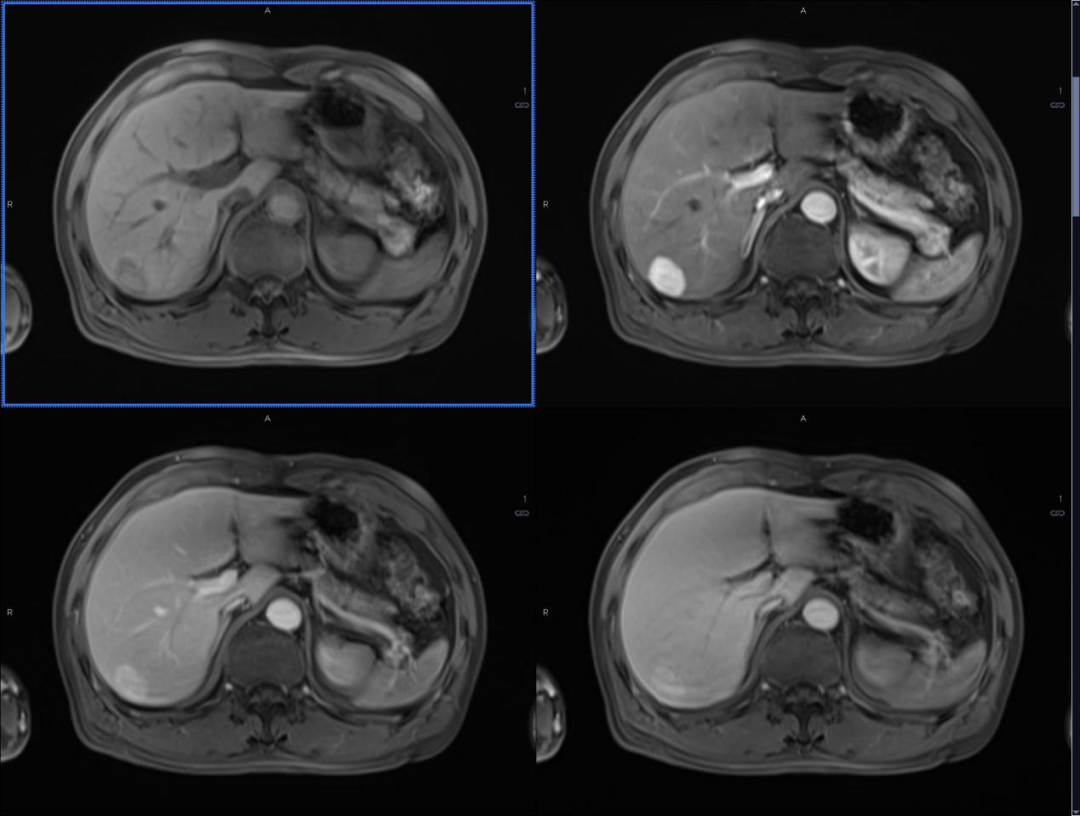
Image produced by Gd-EOB-DTPA enhanced MRI
The result of enhanced MRI showed that there was a lump in the upper right side of the liver (Segment 7). In radiologists’ discussion, it came down to a FNH.
The family was told that FNH was a relatively benign lesion requiring regular visit. They all wept with joy. But they still wanted it resected.
The pathology report a week after the surgery confirmed it was FNH. The family was satisfied and relieved. In consideration of her hepatitis B history, regular liver Doppler ultrasound, alpha-fetoprotein and liver function test were suggested.
Health Tips
A lump in the liver involves a variety of factors and often manifests no specific symptoms itself. Imaging is essential to the detection and pathology is the litmus test. So do not rush to the worst-case-scenario thinking in this situation. Seek professionals that make diagnosis based on manifestations of imaging result, differentiation of tissues, presence of involvement and count of lesions.
Gd-EOB-DTPA enhanced MRI is a good choice in the case of liver tumor smaller than 1cm. It elevates early diagnosis rate of liver tumor and reveals the nature quickly.