
Recently, Ms. Chen (pseudonym) underwent further examination accompanied by her family. There were four people in total, each displaying an anxious and nervous expression on their faces.
After the MRI with Primovist® contrast enhancement, it was found that there was a mass in segment S7 of the right liver lobe. After comprehensive discussion, the radiologist considered it to be focal nodular hyperplasia (FNH) of the liver.
After understanding the situation, the radiologist learned that Ms. Chen had occasional abdominal pain over the past year, which she had attributed to gastric issues and treated with over-the-counter medications. However, this year, due to more frequent abdominal pain, she underwent ultrasound and CT scans at a local hospital, revealing a liver mass. Given the patient's history of hepatitis B, the doctor diagnosed a high possibility of liver cancer.
This news frightened Ms. Chen and her family. How could such a healthy person suddenly develop liver cancer? Unable to accept this sudden and distressing news, Ms. Chen decided to seek advice from specialists in Guangzhou.
Eventually, through a referral, Ms. Chen and her family came for further examinations to clarify the situation. It was this examination that brought a new hope to Ms. Chen and her family.
Based on the CT scan results from the previous hospital, the radiologist recommended an MRI with Primovist® contrast enhancement.

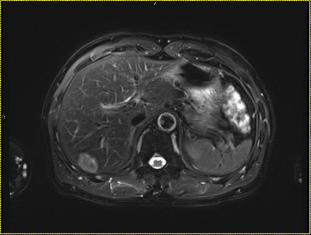
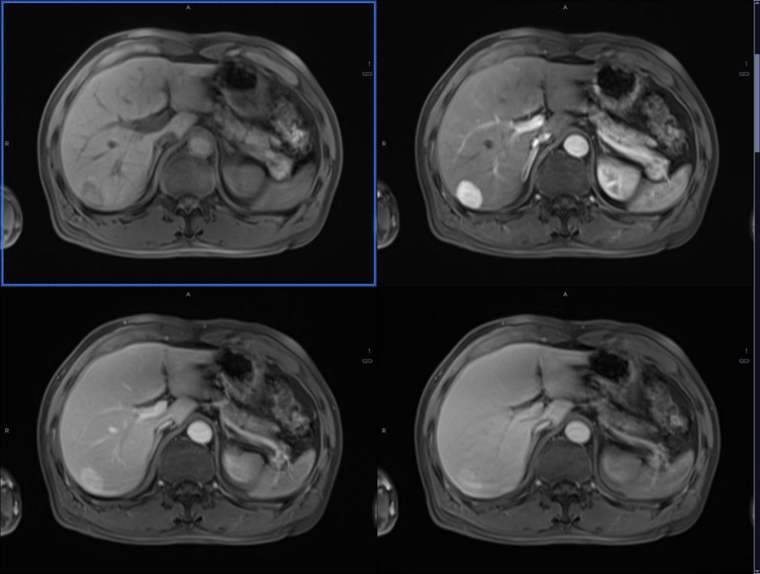
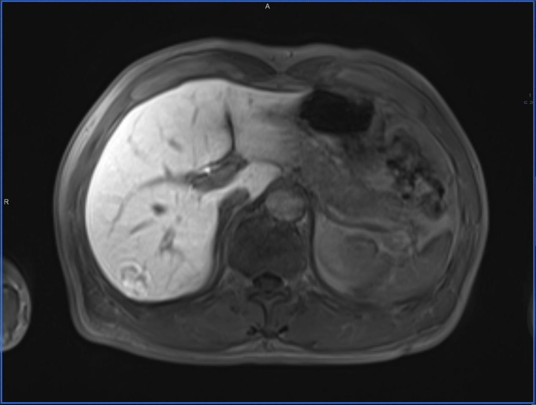
Royal Lee Cancer Center MRI with Primovist® contrast enhancement imaging data
Patient History: Female, 28 years old, with a history of hepatitis B for over ten years; AFP: 55.3.
Ultrasound from the previous hospital showed a space-occupying lesion in the right anterior lobe, and CT enhancement suggested primary liver cancer in segment S7.
Recently, MRI with Primovist® contrast enhancement revealed a lesion in segment S7 of the liver, with low signal on T1WI, slightly high signal on T2WI, high signal on DWI, and dynamic enhancement showing rapid wash-in and wash-out, with the lesion showing hypointensity in the hepatobiliary phase.
After the MRI with Primovist® contrast enhancement, it was found that there was a mass in segment S7 of the right liver lobe. After comprehensive discussion, the radiologist considered it to be focal nodular hyperplasia (FNH) of the liver.
The radiologist informed the patient's family that FNH is a type of benign liver lesion that only requires regular follow-up checks. Ms. Chen's family burst into tears of joy upon hearing this news. However, due to the family's concerns, they still wished for surgical removal in one go.
After a week of surgery, the pathological results were consistent with the diagnosis of FNH made by MRI with Primovist® contrast enhancement. The patient's family was very satisfied, and their lingering worries were finally put to rest. However, considering the patient's history of hepatitis B, the radiologist recommended regular follow-up liver ultrasound, AFP, and liver function tests.