
As one of the most common malignant tumors in women, breast cancer seriously endangers women's physical and mental health. With the use of comprehensive treatment methods, breast cancer has become one of the most effectively treatable solid tumors. This relies on the popularization of breast cancer screening, especially early screening.
The Importance of Breast Cancer Screening
In clinical practice, due to the lack of typical symptoms and signs in early breast cancer, it is not easily noticed by patients, leading to the progression of the disease. Most patients are discovered during physical examinations or breast cancer screenings.
Generally, typical signs of breast cancer appear in the middle and late stages of cancer, such as nipple lumps, nipple discharge, changes in the skin, abnormal nipples or areolas, and enlarged lymph nodes in the armpit. If there are suspected symptoms in daily life, it is recommended to seek medical attention for examination as early as possible.
Preventing breast cancer, early regular screening is very important!
Breast cancer screening refers to the identification and detection of precancerous lesions and early invasive cancer patients with progression potential among asymptomatic women through effective, simple, and economical breast examination measures, aiming for early detection, early diagnosis, and early treatment. Its ultimate goal is to reduce the mortality rate of breast cancer in the population. It is particularly emphasized that the screening age for high-risk breast cancer populations should be before the age of 40.
High-risk Breast Cancer Populations
1) Those with a clear genetic predisposition to breast cancer;
2) Patients with a history of breast ductal or lobular atypical hyperplasia or ductal carcinoma in situ;
3) Those who have undergone chest radiation therapy.
Common Methods for Breast Cancer Screening
Currently, common breast imaging examinations mainly include mammography, breast ultrasound, and breast MRI.
1. Mammography
The most basic examination method, it has an irreplaceable advantage in detecting calcifications compared to other imaging methods. However, it shows poor display of dense breasts and lesions near the chest wall and has radiation hazards. For women under 40 years old without clear risk factors for breast cancer or no abnormalities found during clinical examination, mammography is not recommended as the first-line examination.
Indications:
- Screening for asymptomatic populations and diagnostic patients:
(1) Screening for asymptomatic populations;
(2) Breast examination for age-appropriate women or those with other relevant abnormalities discovered during screening or other examinations;
(3) Symptoms such as breast masses, local thickening, abnormal nipple discharge, breast skin abnormalities, local pain, or swelling;
(4) Short-term follow-up of benign lesions;
(5) Follow-up after breast-conserving surgery for breast cancer;
(6) Follow-up after breast reconstruction surgery;
(7) Guidance for localization and biopsy.
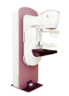
Fully Digital Mammography Machine in Our Hospital
2. Breast Ultrasound
Due to its simplicity, flexibility, intuitiveness, non-invasiveness, and lack of radiation, it is suitable for all populations suspected of breast lesions.
Indications:
(1) Those with breast-related symptoms: palpable breast masses, nipple discharge, inverted nipples, local skin changes, etc.;
(2) Breast examination for asymptomatic high-risk breast cancer populations;
(3) As a supplementary examination for mammography screening;
(4) Follow-up of benign breast lesions; follow-up after breast cancer surgery; follow-up after menopausal hormone replacement therapy, etc.;
(5) Interventional ultrasound: ultrasound-guided fine needle/core needle biopsy and preoperative localization, etc.
3. Breast MRI Examination
Its advantages lie in its high sensitivity, ability to display multiple lesions, multicentric or bilateral breast cancer lesions, and simultaneous display of the relationship between tumors and the chest wall, axillary lymph node metastasis, etc., providing more reliable evidence for formulating surgical plans. However, its specificity is moderate, with a high false-positive rate, unsatisfactory display of tiny calcified lesions, long examination time, and high cost. It is not the preferred examination method.
Indications:
(1) Difficulties in lesion detection or diagnosis with mammography and ultrasound;
(2) Preoperative staging of breast cancer and screening for contralateral breast tumors;
(3) Evaluation of neoadjuvant chemotherapy efficacy;
(4) Identification of the primary lesion in patients with axillary lymph node metastasis;
(5) Differentiation between post-treatment scars and tumor recurrence after breast cancer surgery;
(6) Evaluation of residual lesions in patients with positive margins after tumor excision;
(7) Evaluation after breast implantation surgery;
(8) Breast cancer screening for high-risk populations;
(9) Guidance for localization and biopsy of breast lesions.